Introduction
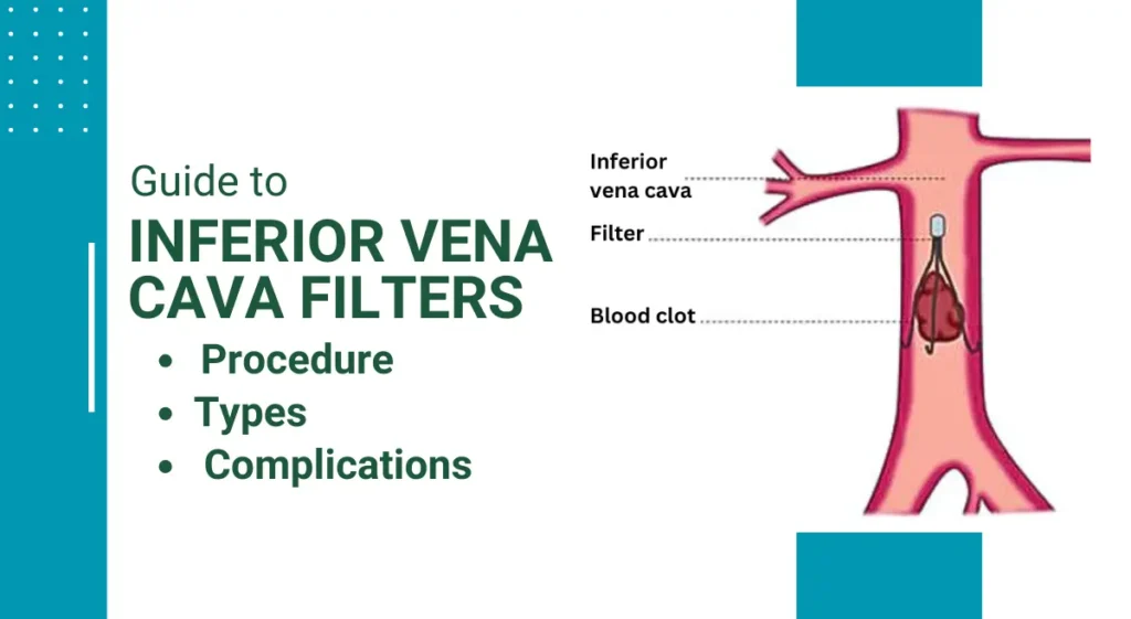
Inferior vena cava (IVC) filters serve as vital tools in the prevention of life-threatening pulmonary embolism by intercepting blood clots and preventing their migration to the lungs. This comprehensive guide provides an in-depth exploration of the uses, types, insertion procedure, complications, and considerations associated with IVC filters, offering valuable insights for healthcare professionals and patients alike.
Table of Contents
Uses of Inferior Vena Cava Filters
- Prevention of Pulmonary Embolism: IVC filters play a crucial role in preventing pulmonary embolism by trapping blood clots within the inferior vena cava, thereby reducing the risk of embolic events.
- High-Risk Patients: Patients with a history of deep vein thrombosis (DVT), pulmonary embolism, or those unable to tolerate anticoagulant therapy may benefit from IVC filter placement to mitigate the risk of recurrent thromboembolic events.
- Prophylactic Use: IVC filters may be employed prophylactically in high-risk surgical or trauma patients to prevent venous thromboembolism during periods of immobility or increased thrombotic risk.
Types of Inferior Vena Cava Filters
- Retrievable Filters: Temporary IVC filters are designed for short-term placement and subsequent retrieval, offering flexibility and adaptability for patients requiring temporary protection.
- Permanent Filters: Intended for long-term placement in patients at risk of recurrent pulmonary embolism, permanent IVC filters provide ongoing protection without the option for retrieval, catering to individuals with contraindications to anticoagulant therapy.
Step-by-Step Guide to Inserting IVC Filters: Detailed Procedure Insights
Patient Preparation:
- The patient is positioned on the procedural table, and the insertion site (typically the femoral or jugular vein) is meticulously cleansed and draped in a sterile fashion.
Vascular Access:
- Utilizing ultrasound guidance, a vascular access sheath is introduced into the vein to facilitate catheter placement and subsequent filter deployment.
Catheter Insertion:
- A catheter incorporating an attached IVC filter is advanced through the vascular sheath under fluoroscopic guidance, precisely guided to the desired location within the inferior vena cava.
Deployment:
- Once the catheter is optimally positioned, the IVC filter is deployed, expanding to create a mesh-like scaffold within the vena cava, effectively capturing and retaining blood clots.
Imaging Confirmation:
- Fluoroscopy or intravascular ultrasound is employed to confirm the accurate placement of the IVC filter and assess for any immediate complications or malpositioning.
Removal of Catheter:
- Upon successful deployment of the filter, the catheter and sheath are carefully withdrawn, and gentle pressure is applied to the insertion site to achieve hemostasis.
Post-procedure Monitoring:
- Following the intervention, the patient undergoes vigilant monitoring for potential complications, and comprehensive post-procedural care instructions are provided.
Complications and Considerations
- Thrombosis: IVC filters carry a risk of thrombosis, encompassing both filter-related thrombosis and thrombotic events occurring at the site of filter insertion, necessitating diligent surveillance and prompt intervention.
- Migration and Embolization: While uncommon, IVC filters may migrate from their intended position or embolize to distant anatomical sites, potentially precipitating serious complications such as organ perforation or vessel obstruction.
- Perforation and Fracture: Prolonged indwelling IVC filters may be associated with vessel perforation, filter tilt, or fracture, posing challenges for retrieval and potentially necessitating surgical intervention.
- Retrieval Challenges: Retrieval of retrievable IVC filters may be complicated by factors such as filter adherence to the vessel wall, significant tilt, or incorporation into the vascular endothelium, necessitating advanced retrieval techniques or adjunctive procedures.
Conclusion
A comprehensive understanding of the indications, types, insertion procedure, complications, and considerations associated with IVC filters is imperative for healthcare providers engaged in the management of venous thromboembolic disease. By adhering to standardized insertion techniques, vigilantly monitoring for complications, and addressing retrieval challenges when encountered, clinicians can optimize patient outcomes and mitigate potential adverse events in this critical aspect of VTE prophylaxis and management.
Commonly Asked Questions about "Inferior Vena Cava Filters"
- An Inferior Vena Cava (IVC) filter is a medical device designed to prevent blood clots from reaching the lungs, where they can cause pulmonary embolisms. It works by trapping blood clots in the inferior vena cava, the large vein that carries blood from the lower body to the heart, thus preventing them from reaching the lungs.
- IVC filters are typically recommended for patients at high risk of pulmonary embolism who cannot take anticoagulant medications or who have experienced recurrent blood clots despite anticoagulation therapy. They may also be used prophylactically in certain high-risk surgical or trauma patients.
- Yes, there are two main types of IVC filters: retrievable and permanent. Retrievable filters are designed for temporary use and can be removed once the risk of pulmonary embolism has passed. Permanent filters, on the other hand, are intended for long-term placement and remain in the body indefinitely.
- The insertion of an IVC filter involves a minimally invasive procedure performed by an interventional radiologist or vascular surgeon. A catheter is inserted into a vein in the groin or neck and guided to the inferior vena cava under imaging guidance. The filter is then deployed, expanding to form a mesh-like structure within the vein.
- Although IVC filters are generally safe, they can be associated with certain risks and complications. These may include thrombosis (blood clot formation), migration or embolization of the filter, vessel perforation, and difficulty in filter retrieval.
Yes, retrievable IVC filters can typically be removed once they are no longer needed. The retrieval procedure is similar to the insertion procedure and is usually performed by an interventional radiologist or vascular surgeon.
- Recovery after IVC filter insertion is usually quick, with most patients able to resume normal activities shortly after the procedure. However, it is essential to follow any post-procedure instructions provided by the healthcare provider, including restrictions on physical activity and medications.
- Retrievable IVC filters are temporary and can be removed once they are no longer needed. Permanent filters, however, are designed for long-term placement and remain in the body indefinitely.
Studies have shown that IVC filters are effective in reducing the risk of pulmonary embolism in high-risk patients. However, their effectiveness may vary depending on factors such as patient selection, filter type, and adherence to post-procedure care.
- During the insertion procedure, patients can expect to receive local anesthesia at the insertion site to minimize discomfort. The procedure itself is typically performed in a specialized interventional radiology suite or catheterization laboratory and may take about an hour to complete. After the procedure, patients are usually monitored for a brief period before being discharged home.